August 15, 2018 feature
Revealing the retina: Graphene corneal contact lens provides robust, irritation-free topographic electroretinography

Our vision can be damaged or lost by damage to the retina—a sensory membrane lining the back of the eye that senses light, converting the image formed into electrochemical neuronal signals—resulting from two classes of medical conditions: a number of inherited degenerative conditions—including retinitis pigmentosa, Leber's congenital amaurosis, cone dystrophy, and Usher Syndrome—as well as diabetic retinopathy, central retinal vein occlusion, sickle cell retinopathy, toxic an autoimmune retinopathies, retinal detachment, and other ocular disorders. To be properly diagnosed and treated (especially when a cataract compromises ophthalmoscopy, 2-D fundus photography, 3-D optical coherence tomography, and other retinal imagery tools), such medical conditions rely on electroretinography—a sensitive technique that detects and measures electrical potential changes at the eye's corneal surface produced in response to a light stimulus by neuronal and non-neuronal retinal cells. Nevertheless, electroretinography has historically faced challenges in the ocular interface electrodes needed to detect an electroretinogram (ERG), these being patient discomfort due to hard electrodes, limited types of electroretinograms with a single type of electrode, reduced signal amplitudes and stability, and excessive eye movement. Recently, however, scientists at Peking University, Beijing, have demonstrated soft, transparent GRAphene Contact lens Electrodes (GRACEs) for conformal full-cornea electroretinogram signal recording in rabbits and cynomolgus monkeys, showing that their soft graphene contact lens electrodes address these limitations.
Prof. Xiaojie Duan discussed the paper that she, graduate students and lead authors Rongkang Yin and Zheng Xu, and their co-authors published in Nature Communications. The biggest challenge in fabricating soft graphene contact lens electrodes with broad-spectrum optical transparency, Dr. Duan told Phys.org, was fabricating wrinkle-free contact lens electrodes, explaining that wrinkles can cause optical inhomogeneity across the electrode, thereby affecting ocular refraction and the accuracy of the light stimulus pattern on the retina. "This in turn undercuts retinopathy diagnosis efficacy," Dr. Duan added. "Graphene obtained from conventional growth method is a flat film, and wrinkles inevitably form after transferring the flat graphene film to the curved surface. To make a graphene contact lens electrode with high electrical conductivity and optical uniformity across the electrode, it's important to directly use a curved graphene film with uniform thickness."
Applying GRACEs to conformal full-cornea electroretinographic recording presented no major obstacles, she continued. "While there's no primary difficulty in applying GRACEs to conformal and full-cornea electroretinographic recording as long as the fabricated GRACEs have reasonable impedance and optical transparency, we can always record high-quality ffERG and mfERG signals. Therefore, to get GRACES with reasonable impedance and optical transparency, graphene film with sheet resistance"—a measure of the resistance of thin films that are nominally uniform in thickness—"below 2000 Ω/sq and optical transparency above 70% will be good enough."
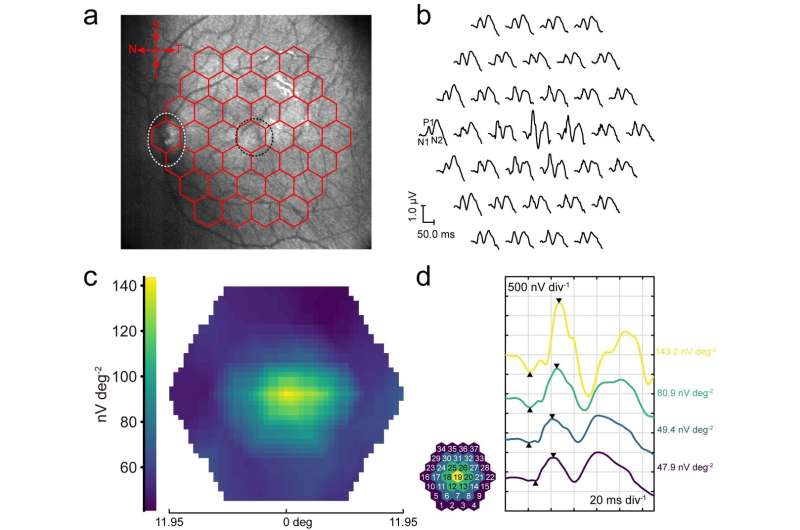
However, the leading challenge for general ERG recording is to measure multifocal ERG (mfERG)—which simultaneously measures local retinal responses from up to 250 retinal locations within the central 30 degrees mapped topographically—reflects the retinal response to stimulation on a specific small retinal area. "For multifocal ERG measurements," Dr. Duan told Phys.org, "the light stimulation pattern is projected onto the retina. It is therefore important for the eye to have proper refraction so that the stimulus pattern can be projected clearly." In addition, the signal amplitude of multifocal ERG is only about 1/1000th that of conventional full-field ERG (ffERG, which measures ERG under entire retina stimulation with a light source under scotopic (dark-adapted) or photopic (light-adapted) retinal adaptation), while mfERG requires relatively longer recording period—making sensitivity, comfort, and stable interfacing with the eye very critical for multifocal ERG recording. "Conventional contact lens electrodes tend to alter the eye's refraction," she pointed out, "which makes them unsuitable for multifocal ERG recording." That said, other electrodes (for example, DTL electrodes), will not alter the eye's refraction but suffer from low measurement sensitivity and signal stability.
Another consideration, Dr. Duan noted, is that the spatial distribution of the ERG potential across the cornea has been a long-existing question. "Conventional electrodes use opaque metal as recording elements, which can only be located at the periphery of the cornea in order to avoid blocking vision—a situation that prevents multi-site spatially-resolved ERG recording, which is necessary to reveal the ERG potential distribution across the cornea. Another factor is that for a conventional stiff electrode, there is always thick tear film between the electrodes and the cornea, which can shunt the potential difference between different locations on the cornea." This last issue introduces yet another significant challenge in elucidating the ERG spatial distribution across the cornea.

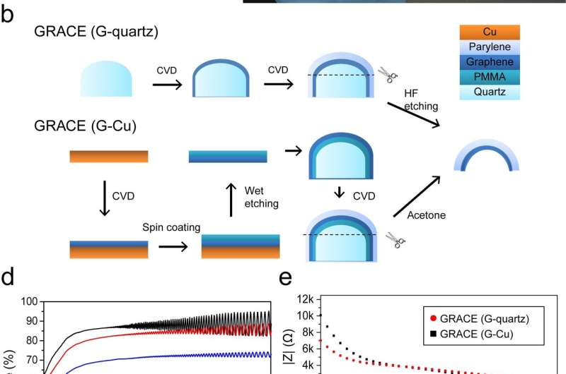
Dr. Duan described the key insights, innovations and techniques they leveraged to address these challenges. "As I mentioned, we eliminate wrinkles by using a curved graphene film directly grown on curved quartz mold—and the film's shape and curvature can be easily tuned by changing those of the quartz molds." The key point, she emphasizes, is the curved graphene film's uniform thickness leads to the resulting GRACEs having uniform electrical conductivity and optical transparency across the entire contact lens electrode, which is what is unique about the team's GRACEs when compared to previously-reported graphene-based eye interfacing devices. "In addition," she added, "we established and optimized the electrode fabrication flow." She emphasizes that by directly depositing ultrathin insulating film (Parylene-C, which forms the GRACE substrate) onto the graphene/quartz complex, and then etching the quartz mold, GRACE devices can be readily fabricated." The key takeaway is that this fabrication strategy avoids poly(methyl methacrylate) (PMMA)—a transparent thermoplastic (also referred to as an acrylic or acrylic glass) commonly used for graphene transfer, which not only avoids possible PMMA contamination that could cause optical inhomogeneity, but also maintains graphene film integrity—a factor critical to maintaining GRACE electrical conductivity.
As previously noted, it is challenging to record multifocal ERG signals with contact lens electrodes because it tends to alter ocular refraction. "To solve this problem," Dr. Duan pointed out, "we designed the GRACE to be soft and conformable to the cornea surface with a tight GRACE/cornea interface." This avoids the formation of thick liquid gaps or air gaps between the electrode and the cornea—the main origin of refraction change when wearing hard contact lens electrodes. As shown in their paper, GRACEs can successfully record high-quality multifocal ERG signals, which is indicative of the advantages of GRACEs over hard contact lens electrodes.
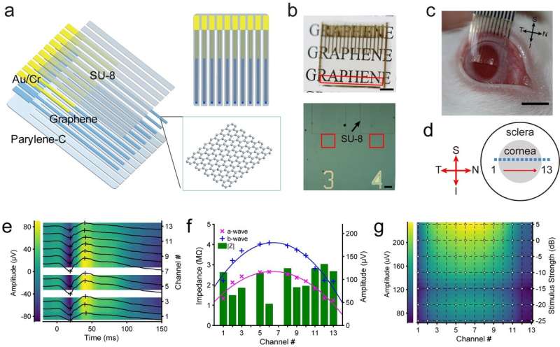
To provide efficient multi-site, spatially-resolved ERG recording, the scientists designed and deployed a soft, transparent graphene multi-electrode array. "The soft electrode's tight interface with the cornea avoided tear film shunting," she explained, "and high optical transparency enables placement of high-density electrode array across the entire corneal surface without blocking the vision or affecting the light stimulus uniformity." As a result, they observed a stronger signal at the central cornea than the periphery, proving the advantages of the soft transparent graphene-based electrodes in ERG recordings.
As to implications of their findings regarding GRACE for in vivo visual electrophysiology studies, Dr. Duan reiterated that their graphene-based contact lens electrodes show the capability for high-efficacy recording of various kinds of ERG recording, including ffERG, mfERG, and meERG (multi-electrode ERG, which maps spatial differences in retinal activity using a conventional full-field stimulus and an array of electrodes on the cornea)—a flexibility not achievable by conventional ERG electrodes. "With further testing and development," she underscored, "it could replace the traditional electrodes and be used in clinical practice. In addition, because retinal lesions can cause change of the local corneal potentials, the multi-electrode ERG recording with the graphene microelectrode array demonstrated herein provides a potential functional retinal electrophysiological imaging technique that can be used as a diagnostic tool for detecting local areas of retinal dysfunction under single full-field stimulus."

Moving forward, Dr. Duan identified three planned next steps in the scientists' research, these being:
- Improving electrode gas permeability to make it more suitable for long-term wear
- Fabricate high-density two-dimensional soft transparent electrode array to map the ERG potential across the entire corneal surface
- Apply the soft transparent graphene microelectrode array for in vivo recording of electrical activity of retinal ganglion cells at single-cell level
She also discussed research and other innovations they might consider developing. "Based on nanomaterials and nanotechnology, we seek to develop techniques that can record or modulate neural activities at large scales with high spatio-temporal resolution and long-term stability, and to explore the application of these techniques in understanding fundamental and pathological brain processes."
In closing, Dr. Duan listed other areas of research that might benefit from their study. "Soft transparent electrodes also enable simultaneous electrophysiology and optical neural imaging or stimulation, which is important for studying the connectivity and function of neural circuits. Conventional neural surface electrode arrays using opaque metal conductors are not suitable for use in simultaneous electrical and optical neural interfacing because they block the field of view and are prone to producing light-induced artifacts in the electrical recordings. The soft transparent graphene microelectrode array described herein can be used in research combining optical and electrical modalities in neural interfacing."
More information: Soft transparent graphene contact lens electrodes for conformal full-cornea recording of electroretinogram, Nature Communications Volume 9, Article number:2334 (2018), https://doi.org/10.1038/s41467-018-04781-w.
Journal information: Nature , Nature Communications
© 2018 Phys.org


