Related topics: cells · cancer cells · protein

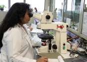
Research team resolves decades-long problem in microscopy

When viewing biological samples with a microscope, the light beam is disturbed if the lens of the objective is in a different medium than the sample. For example, when looking at a watery sample with a lens surrounded by ...

Discovery of the first fractal molecule in nature

An international team of researchers led by groups from the Max Planck Institute in Marburg and the Philipps University in Marburg has stumbled upon the first regular molecular fractal in nature. They discovered a microbial ...

page 1 from 40

Electron microscope

An electron microscope is a type of microscope that uses a particle beam of electrons to illuminate a specimen and create a highly-magnified image. Electron microscopes have much greater resolving power than light microscopes that use electromagnetic radiation and can obtain much higher magnifications of up to 2 million times, while the best light microscopes are limited to magnifications of 2000 times. Both electron and light microscopes have resolution limitations, imposed by the wavelength of the radiation they use. The greater resolution and magnification of the electron microscope is because the wavelength of an electron; its de Broglie wavelength is much smaller than that of a photon of visible light.

The electron microscope uses electrostatic and electromagnetic lenses in forming the image by controlling the electron beam to focus it at a specific plane relative to the specimen. This manner is similar to how a light microscope uses glass lenses to focus light on or through a specimen to form an image.

This text uses material from Wikipedia, licensed under CC BY-SA