Cell behavior, once shrouded in mystery, is revealed in new light

A cell's behavior is as mysterious as a teenager's mood swings. However, University of Missouri researchers are one step closer to understanding cell behavior, with the help of a specialized microscope.
Previously, in order to study cell membranes, researchers would often have to freeze samples. The proteins within these samples would not behave like they would in a normal biological environment. Now, using an atomic force microscope, researchers can observe individual proteins in an unfrozen sample—acting in a normal biological environment. This new observation tool could help scientists better predict how cells will behave when new components are introduced.
"What's missing right now in cell biology is the ability to predict cell behavior," said Gavin King, associate professor of physics and astronomy in the MU College of Arts and Science, and joint assistant professor of biochemistry. "We don't know all of the details yet on a number of biological processes. For example, when a drug is introduced to a cell, it must pass through the membrane, which may create a reaction. The more knowledge we have about that reaction, the better we will be able to create drugs that can target a specific area and, possibly, result in fewer side effects."
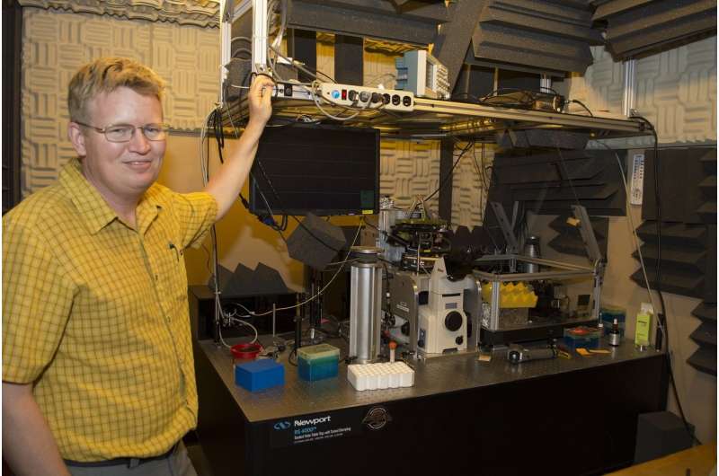
The atomic force microscope is capable of tracing the three-dimensional shape of an individual protein in biological conditions (in fluid at room temperature). It is comprised of a robotic arm with a tiny needle attached on one end. Researchers position the arm precisely on the sample they wish to analyze. Then, by very gently tapping the needle multiple times into the specimen in various points, a real-time, three-dimensional image of a protein is developed.
For this study, researchers focused on imaging the consequences of a chemical reaction occurring within one particular protein from E. coli that is responsible for transporting other proteins across the cell membrane. They picked E. coli for this study because of the simplicity of its cells. While researchers could not control the precise moment the reaction occurred, the force microscope's tapping motion allowed researchers to watch in real time how that protein changed its shape in response to the release of chemical energy. These conformational changes are directly related to the protein's biological function.
"We can keep our eyes on just one protein, add various components, and then watch what happens," King said. "It is like making a movie of a single molecule doing its biological work. We are really in the early days of understanding the mechanical details of how cells work, but as these tools become increasingly more precise they could provide us with essential information in the future."
The study, "Single molecule observation of nucleotide induced conformational changes in basal SecA-ATP hydrolysis," was published in Science Advances.
More information: Nagaraju Chada et al. Single-molecule observation of nucleotide induced conformational changes in basal SecA-ATP hydrolysis, Science Advances (2018). DOI: 10.1126/sciadv.aat8797
Journal information: Science Advances
Provided by University of Missouri-Columbia


